‘Eye-Opening’ Study Sheds Light on Circadian Pacemaker
University of Maryland researchers studied synaptic changes in an animal model before and after it developed vision, which plays a key role in circadian rhythm.
In a new study featured on the cover of the journal Analytical Chemistry on October 17, 2023, a team of University of Maryland researchers used cutting-edge technologies to study a little-understood visual pathway in an animal model.
The research team set out to understand how the development of vision affects proteins in a part of the brain called the suprachiasmatic nucleus (SCN), which is considered the main circadian pacemaker in mammals because of its ability to regulate circadian rhythms in response to light.
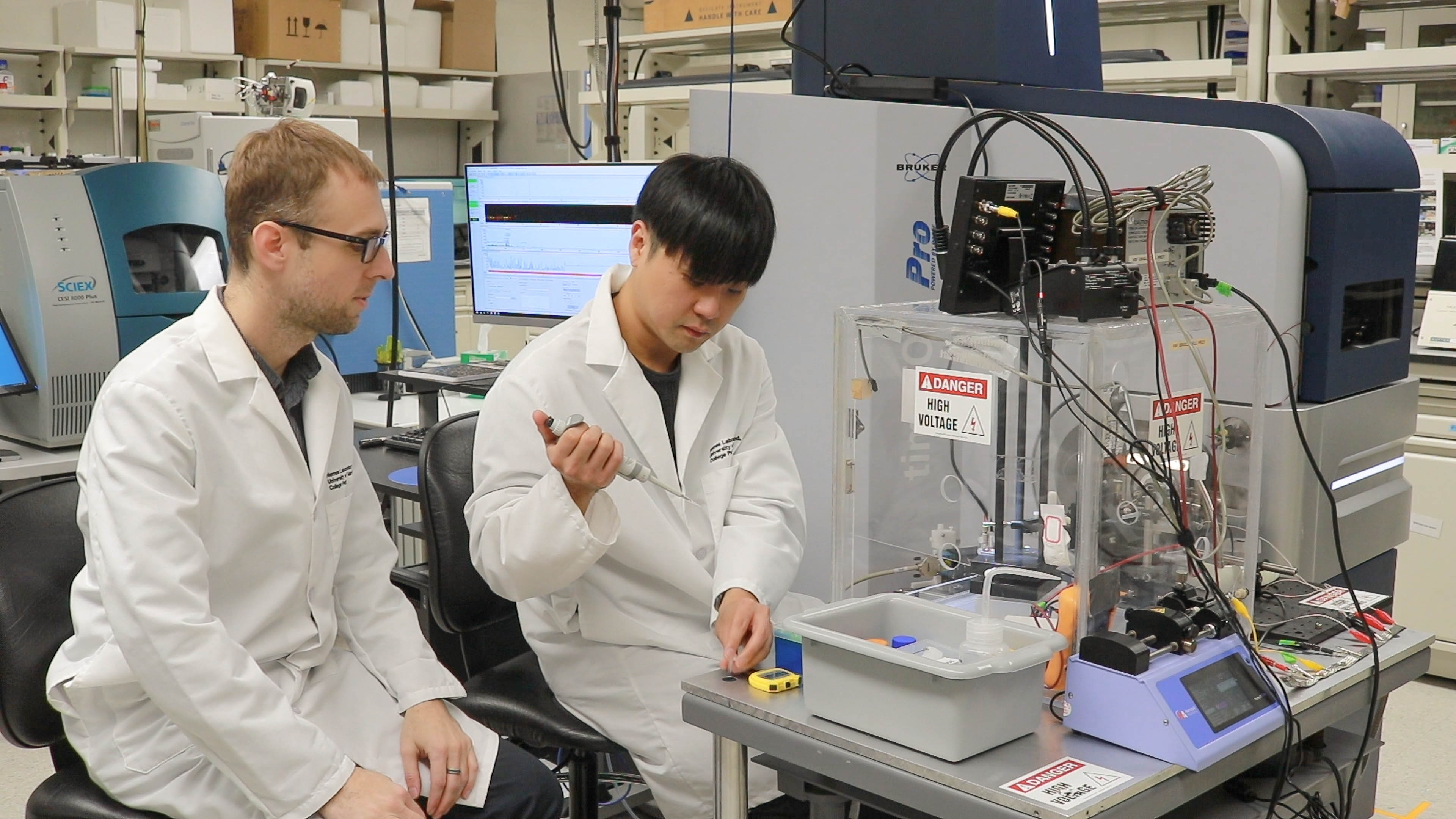
Designed by Peter Nemes, a professor in the Department of Chemistry and Biochemistry, and Colenso Speer, an assistant professor in the Department of Biology, the study stems from a 2019 seed grant from UMD’s Brain and Behavior Institute (BBI) that also included Cell Biology and Molecular Genetics Professor Najib El-Sayed. Nemes previously received a Beckman Young Investigator award from the Arnold and Mabel Beckman Foundation in 2015, which enabled the construction of the mass spectrometer that Nemes further refined at UMD for this latest project.
Their Analytical Chemistry study fused two advanced and disparate technologies: mass spectrometry and microscopy.
“The BBI award was so important for Colenso and me because it allowed us to get together and integrate these totally different disciplines,” Nemes said. “I work in chemistry with mass spectrometry, which can identify and quantify up to a thousand different proteins in a neuron. With high-resolution microscopy, Colenso can zoom into tissue—down to the level of single cells, axons and dendrites—and provide exquisite spatial resolution.”
With the ability to pinpoint where synapses are forming, the research team determined that various proteins increased in production when a mouse opened its eyes for the first time—an important developmental stage linked to circadian rhythm. Levels of a protein called synapsin spiked after eye opening due to the growth of new synapses, but they did not come from the retina. Instead, they likely originated in the hypothalamus—the region of the brain that houses the circadian pacemaker.
“These findings tell us that there are different mechanisms of synaptic organization and development in distinct visual circuits critical for behavior and physiology,” Speer said. “Understanding these differences in synaptic specificity and development is a critical step toward understanding the many ways that light regulates brain function outside of what we normally think of as vision.”
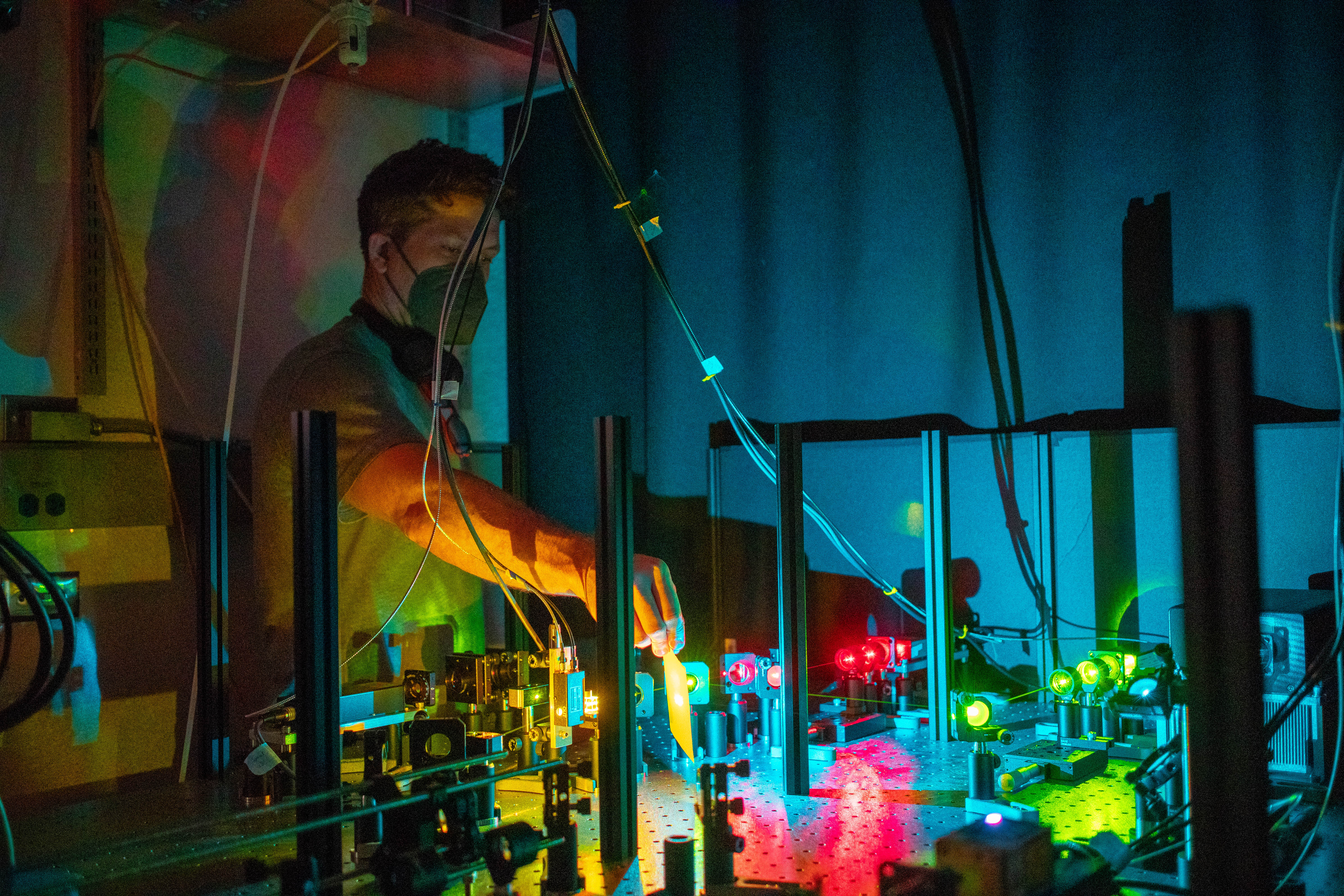
Their study is the first to identify proteins and image synapses in a mouse’s SCN at such an early developmental stage. While they focused on a specific visual pathway, Nemes and Speer said that researchers have a broader interest in understanding the circadian pacemaker, in part because of its importance to human health.
“There are human health concerns with circadian physiology, most notably shift work disorder,” Speer said. “A common one that we all suffer from at some point is jet lag, which is a disconnect between your body’s intrinsic circadian rhythm and the light cycle of the new location that you find yourself in.”
Speer noted that the technologies he and Nemes developed could be applied to a wide range of studies. In the future, they could even extend to research on human health and disease.
“We could envision opportunities for applying this type of imaging technology and mass spectrometry to biopsied or postmortem samples as preclinical research to help establish the commonalities and differences between human neural circuits and these animal models that we’re working on in the laboratory,” Speer said. “I think this opens up a lot of exciting opportunities in terms of understanding how local proteins in synapses are regulated.”
Nemes is excited to see how their blended technologies will be applied in future research, adding that their Analytical Chemistry paper is just the beginning.
“By integrating ultra-high-sensitivity mass spectrometry with super-resolution imaging to discover new findings, you could apply this to any other context in the brain,” Nemes said. “This work lays the foundation for many more studies to come.”
###
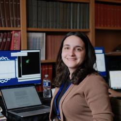
In addition to Nemes and Speer, UMD-affiliated co-authors included Theresa Alexander (B.S. ’15, mathematics; Ph.D. ’23, biological sciences), Sam Choi (B.S. ’10, Ph.D. ’21, chemistry), Shyrice Mitchell (M.S. ’21, biological sciences), neuroscience and cognitive science Ph.D. student Tarlan Vatan (B.S. ’20, biological sciences) and Chenghang Zhang (Ph.D. ’23, biophysics).
Their paper, “Microanalytical Mass Spectrometry with Super-Resolution Microscopy Reveals a Proteome Transition During Development of the Brain’s Circadian Pacemaker,” was published online in Analytical Chemistry on October 4, 2023.
This work was supported by the Arnold and Mabel Beckman Foundation, the Brain and Behavior Research Foundation, the National Institutes of Health (Award Nos. DP2MH125812 and 1R35GM124755), the National Science Foundation (Award No. IOS-1832968), and UMD’s Brain and Behavior Institute. This article does not necessarily reflect the views of these organizations.